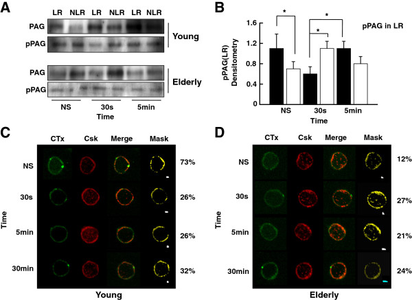
Figure 3.
Western blot and confocal analysis of Csk and tyrosine-phosphorylated PAG in plasma membrane lipid rafts (LR). (A) Purified T cells were left non-stimulated (NS) or exposed to a mixture of anti-CD3 and anti-CD28 (5 μg/ml each) mAbs for various periods of time, as indicated. Cell lysates were separated on sucrose density gradients and fractions corresponding to lipid rafts (LR) and non-lipid rafts (NLR) were isolated, sized by SDS-PAGE under reducing conditions, electrotransfered to nitrocellulose membranes and proteins revealed using appropriate mAbs and the chemiluminescence technique. β-Actin was used as control of gel loading. The protein transferred to nitrocellulose membranes were stained with Ponceau to verify that similar amounts of protein had been loaded in each lane. (B) Protein bands were analyzed by semi-quantitative densitometry and are reported in arbitrary units. Results of T cells of young (filled columns) and elderly (empty columns) donors are shown. Data are represented as the mean ± SD. Asterisks indicate statistical significance (Student’s t-test) for p < 0.05. (C and D) Confocal analysis of the distribution in lipid rafts of cholera toxin B subunit (CTx) and Csk of resting (NS) and T cells activated (TCR-CD28) for various periods of time, as indicated. An illustrative example of young (C) and elderly (D) individual, merged images and masking are shown. Data are representative of one of a minimum of 12 independent experiments.

