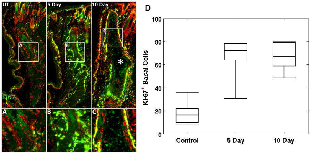
Figure 4.
Ductal dilation and increased Ki67 staining were observed in meibomian glands of dry eye mouse model. Shown in panel A is an overlay of Ki67 (green) and DAPI (red) stained images of meibomian gland from untreated, normal mice. Panel B and C show stained images of glands from mice treated in a desiccating stress environment for 5 and 10 days, respectively. In panel D, the average number of cells that show positive Ki-67+ stain under each categories are shown. Central line marks the median.

