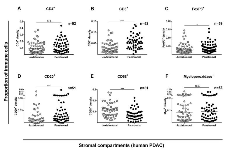
Figure 2. Stromal compartment specific immune cell infiltration in human PDAC.
Proportion of immune cell infiltrate was determined as shown in Supplementary Figure 1-4. Juxtatumoral stromal (within 100μm of tumor cells (single or islands /ducts)) and panstromal (the rest of the tumor stroma) were defined. Each data point represents a single patient (median scores of all TMA cores (n=6)) and lines represent median for the cohort of patients. Comparison of immune cell infiltration in these two PDAC stromal compartments was made for CD4+ (A), CD8+ (B), FoxP3+ (C), CD20+ (D), CD68+ (E) and Myeloperoxidase+ (F) cells, and also for CD3+ and CD56+ cells (Supplementary Figure 5). Whilst PDAC tissues demonstrate a significantly higher density in the juxtatumoral stroma relative to the panstromal compartment for CD68+ cells, the reverse was true for CD8+, FoxP3+ and CD20+ cells with an equal density for CD4+ and Myeloperoxidase+ cells implying a differential immune cell infiltration defect. Mann Whitney U test; p-values are two-tailed.

