
Figure 1.
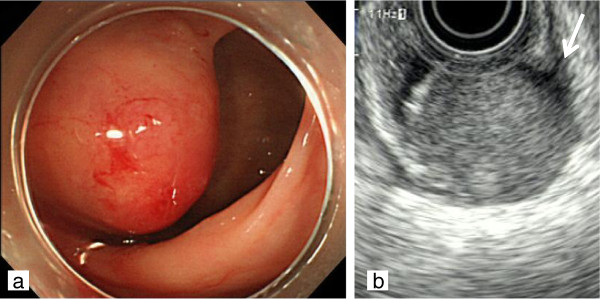
Findings on colonoscopy and endoanal ultrasound scanning. (a) Colonoscopic examination showed a submucosal tumor 3 cm in diameter arising in the posterior rectal wall. (b) Endoanal ultrasound scanning showed a heterogeneous, low-echoic tumor with a nodular appearance arising in the muscularis (long arrow) of the posterior rectal wall.