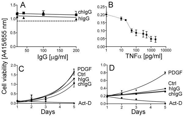
Figure 8.

Analysis of the effects of the chIgG variant on the viability of cells. A; Assays of the viability of dermal fibroblasts as the function of a 24-h treatment with increasing concentrations of the chIgG variant or control hIgG. The dotted line represents data for cells cultured in the absence of antibodies. B; The ability of WST-8-based assays to detect changes in the viability of fibroblastic cells was monitored in dermal fibroblasts cultured in the presence of various concentrations of TNFα. C and D; Proliferation assays of dermal fibroblasts (C) and cells isolated from the tendon sheaths (D) cultured in the presence of 100 μg/ml of tested antibodies. In addition, proliferation of cells cultured in the presence of PDGF or actinomycin D (Act-D) was also monitored. Moreover, a control (Ctrl) group of cells cultured in the absence of tested agents was also analyzed.
