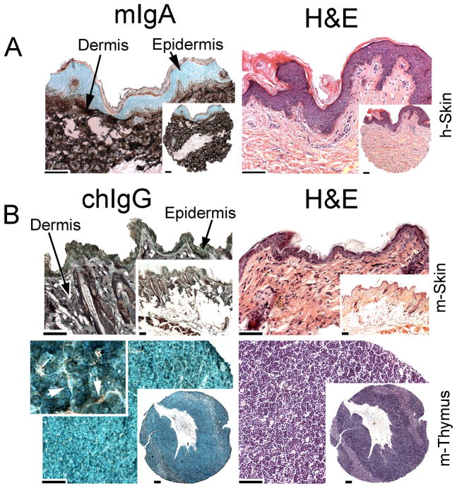
Figure 9.
Cross-reactivity assays of the mIgA and chIgG variants of the anti-α2Ct antibody. The human skin sample (A) was immunostained with the mIgA variant, while the mouse skin and thymus were immunostained with the chIgG form of the anti-α2Ct antibody (B). Corresponding panels were stained with H&E. As depicted in panel A, staining of the human skin (h-Skin) has revealed that collagen I -specific staining was limited to the collagen-rich dermis while the epidermis was collagen I-negative. Panel B demonstrates collagen I-positive staining of the dermal layer of the mouse skin (m-Skin), whereas the epidermis is collagen I-negative. In the mouse thymus (m-Thymus), collagen I-positive staining is apparent only in the interlobular regions (arrows). Bars = 50 μm.

