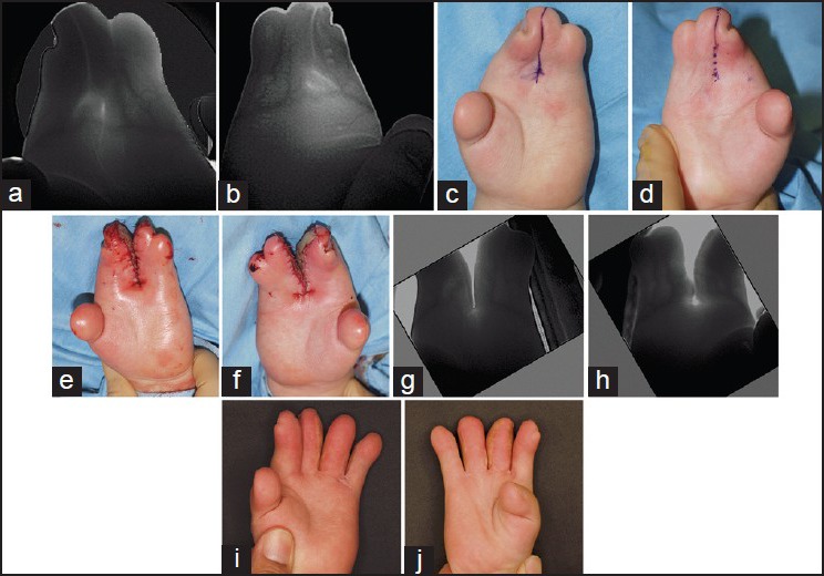
Figure 2.

Infrared venography through finger splitting surgeries for an Apert patient. Volar venous arch placed distally to the metacarpophalangeal joint, where incision had to be performed to divide fingers sufficiently. (a, b) Snap shots of palmar side venogram, immediately before the separation surgery. (c, d) Designs for the first separation surgery, (e and f) Immediately after the first separation surgery. (g, h) Snap shots of venogram, immediately before the second separation surgery. Volar venous arch was severed between the middle and ring fingers. (I, j) Fifteen months after the second separation surgery
