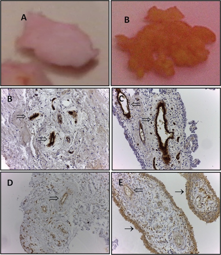
Figure 2.
Macroscopic appearance of N/R (A) and I (B) synovial biopsies. Immunohistochemical detection of von Willebrand’s factor in N/R (C) and I (D) synovial biopsies. N/R and I synovial biopsies were stained with anti-von Willebrand factor antibody. The presented images are representative of the obtained results. Immunohistochemical detection of VEGF in N/R (E) and I (F) synovial biopsies. N/R and I synovial biopsies were stained with anti-VEGF antibody. The presented images are representative of the obtained results. Magnification ×20.
I, inflammatory; N/R, normal/reactive; VEGF, vascular endothelial growth factor; (⇒), blood vessels; (→), intima lining.

