
Figure 4.
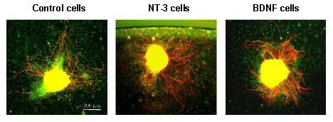
Response of spiral ganglion neurites to cellular sources of neurotrophins.
Explants were immunostained for neurofilament 200 (Texas Red). Non-neuronal cells were visualized with phalloidin (FITC) to label cellular actin to identify any potential relationships between non-neuronal cells and neurites, such as targeting. On a two-dimensional collagen substrate, spiral ganglion explants co-cultured with brain-derived neurotrophic factor (BDNF) secreting cells exhibit enhanced neurite number when compared to explants maintained with control, non-secreting fibroblasts. Neurotrophin-3 (NT-3) secreting cells enhanced neurite length. The images were obtained on an Olympus IX70 inverted fluorescent microscope.