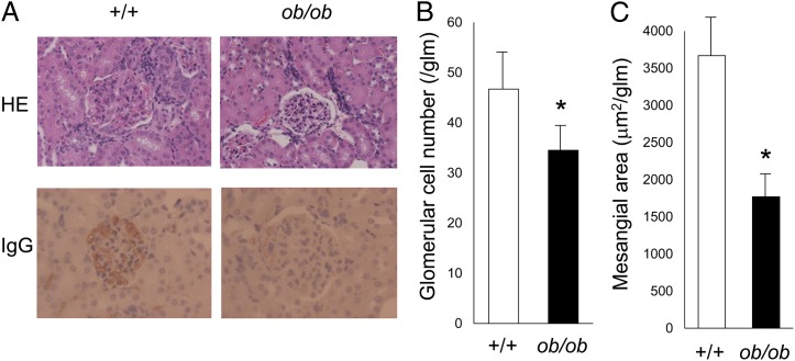
FIGURE 4.
Histological examination of the kidneys of mice with and without deficiencies in leptin signaling. (A) Representative light microscopy images of kidney tissue from 20-wk-old MRL/Mp-Faslpr-ob/ob mice and control littermates. Original magnification ×400. The sections were stained with H&E (upper panels) and Ab to IgG (lower panels). (B) Quantitative analysis of parameters of glomerulonephritis. Glomerular cell number was significantly lower in MRL/Mp-Faslpr-ob/ob mice than in control littermates. Data are mean ± SEM (n = 10/group). (C) Mesangial area within glomerular tufts was significantly lower in MRL/Mp-Faslpr-ob/ob mice than in control littermates. Data are mean ± SEM (n = 10/group). *p < 0.05.

