Figure 4.
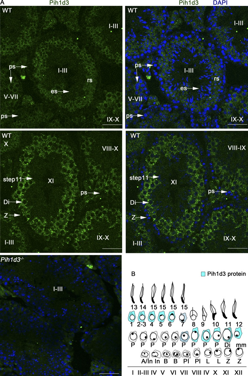
Subcellular localization of Pih1d3 in adult mouse testis. (A) Immunofluorescence staining of the testis of adult WT or Pih1d3−/− mice with antibodies to Pih1d3. Pih1d3 (green fluorescence) was detected in the cytoplasm of WT sperm cells from late pachytene spermatocytes to round spermatids (rs), with its abundance being highest at the diplotene (Di) and pachytene stages. Nuclei were stained with DAPI (blue fluorescence). Stages of the cycle of the seminiferous epithelium are indicated with roman numerals. Images were obtained with a 40× objective lens. Bars, 50 µm. (B) Staging diagram for Pih1d3 protein expression in the adult testis. Circles around the nucleus indicate cytoplasm, with the intensity of the blue color representing the expression level of Pih1d3.

