Figure 1.
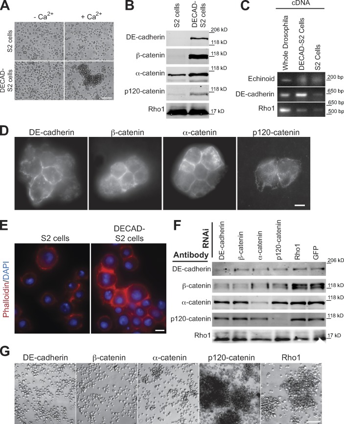
Properties of DECAD-S2 cells. (A) Bright-field microscopy of S2 or DECAD-S2 cells in Schneider’s media or Hank’s buffer with and without Ca2+. (B) Western blots of the indicated proteins in whole-cell lysates of S2 or DECAD-S2 cells. Each lane was loaded with protein from 2.5 × 10⁵ cells. (C) RT-PCR of the indicated genes from cDNA collected from whole Drosophila adults, S2 cells, or DECAD-S2 cells. (D) Immunofluorescence of the indicated proteins in DECAD-S2 cells. (E) Rhodamine phalloidin staining of S2 and DECAD-S2 cells. (F) Western blot analysis of the indicated proteins in different RNAi treatments of DECAD-S2 cells. Each lane was loaded with protein from 2.5 × 10⁵ cells. (B, C, and F) Hashes indicate molecular mass standards. (G) Bright-field microscopy of RNAi-treated cells after plates swirling to induce aggregate formation. Bars: (A and G) 100 µm; (D and E) 2 µm.

