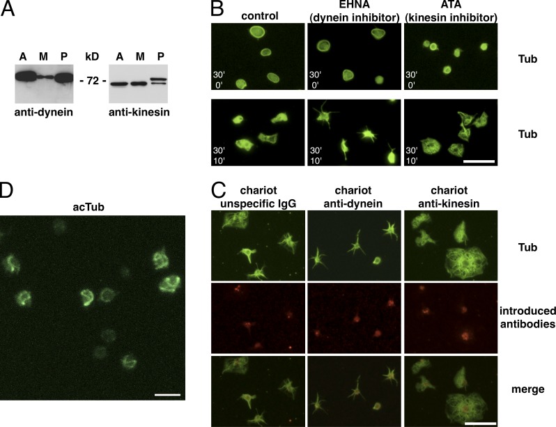
Figure 1.
Presence of microtubule motors in platelets and motor inhibitor treatments. (A) Western blot of 5 µg lysates of the cell line A549 (A), the megakaryocyte precursor line CHRF-288-11 (M), and of 107 platelets (P) revealed with a pan-anti–kinesin heavy chain and an anti–dynein intermediate chain antibody. (B) Resting platelets in PRP from buffy coats were diluted in PBS, 2.5 × 10⁶/ml, and incubated with 1 mM EHNA or 10 µM ATA for 30 min at RT and then either fixed (top; 30’ inhibitors/0’ spreading) or allowed to spread on a glass surface for 10 min (bottom; 30’ inhibitors/10’ spreading) before fixation and α-tubulin staining. (C) Control rabbit IgGs as well as mouse anti-dynein and rabbit anti-kinesin function-blocking antibodies were introduced into living platelets using the Chariot kit. Platelets were then allowed to spread on glass coverslips for 10 min, fixed, and stained using a monoclonal rabbit anti α-tubulin antibody for the anti-dynein Chariot and a mouse anti–α-tubulin antibody for the control and the anti-kinesin Chariot conditions (in green) as well as secondary antibodies recognizing the introduced antibodies (anti–mouse for the dynein Chariot and anti–rabbit for the control and the kinesin Chariot conditions, in red). (D) 3D projection of a confocal z stack of platelets treated as in B (top) with 10 µM ATA but for only 3 min, fixed, and stained for acetylated tubulin (acTub). Video 1. Bars: (B and C) 10 µm; (D) 5 µm.

