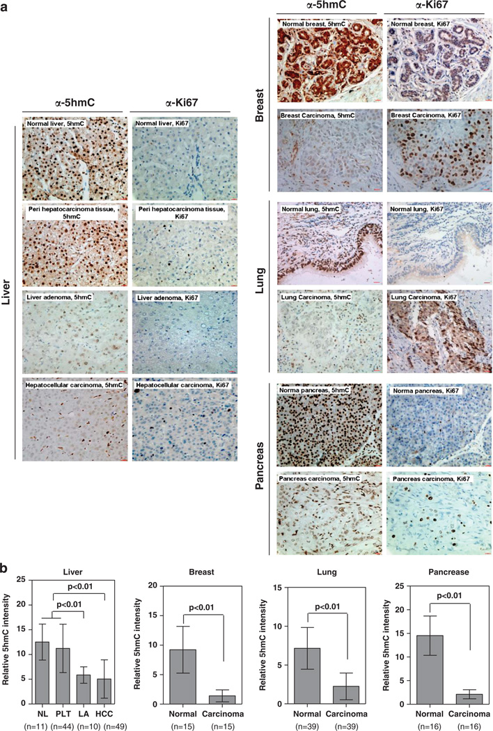
Figure 1.
5hmC is substantially reduced in multiple human tumors. IHC was performed using antibodies against 5hmC and Ki67 in human normal and cancer tissues from liver, breast, lung and pancreas (a). The mean values of the IHC quantification are shown (b). Scale bars are 20 µm. Data are represented as mean±s.d. Freshly frozen or paraffin-embedded human normal tissue and tumors of liver, breast, lung, prostate and pancreas were acquired from Department of Pathology, Huashan Hospital, Fudan University. The procedures relating to the acquisition of samples from human subjects were approved by the Ethics Committee of the Institutes of Biomedical Sciences (IBS), Fudan University. The paraffin-embedded specimens were cut into 5 µm thin sections, which were subsequently stained with hematoxylin and eosin (H/E). Subsequent separation of tumor and normal surrounding tissues were carried out microscopically by an experienced molecular pathologist and guided by the H/E staining. For IHC analysis, the labeled streptavidin-biotin (LSAB) method was applied using commercial kits (Dako Corporation, Santa Barbara, CA, USA). Briefly, paraffin sections were deparaffinized and rehydrated following standard protocols. Sections were incubated with 3% H2O2 to eliminate the endogenous peroxidase activity, and then blocked with 5% normal goat serum. After treatment with 2N HCl for 15 min at room temperature, sections were neutralized with 100mm Tris–HCl (pH 8.5) for 10 min and then washed three times with PBS, followed by incubation with a primary anti-5hmC antibody (Active Motif; Cat. 39769, dilution at 1:1000) at 37 °C for 1 h. A horseradish peroxidase (HRP)-conjugated secondary antibody (Dako Corporation) was then applied and incubated at 37 °C for 1 h. For Ki67 IHC staining, the LSAB method was used as described above, using a primary anti-Ki67 antibody (dilution 1:100; Abcam, Cambridge, MA, USA) and a HRP-conjugated secondary antibody (Dako Corporation). Sections were developed with DAB kit and stopped with water. To semiquantify the 5hmC-positive areas, five randomly selected fields (173 µm2 each) from each sample were randomly selected and microscopically examined. Images were captured using a charge-coupled device (CCD) camera and analyzed using Motic Images Advanced software (version 3.2, Motic China Group Co. Ltd). The relative 5hmC intensity was calculated by dividing the positively stained areas over the total area. All IHC image analysis values were calculated as mean ± s.d. Statistical analysis was performed using SPSS 11.5 statistical package. Statistical methods included Paired t-test and independent samples t-test. All statistical tests were considered significant at an α=0.05 (P < 0.05).

