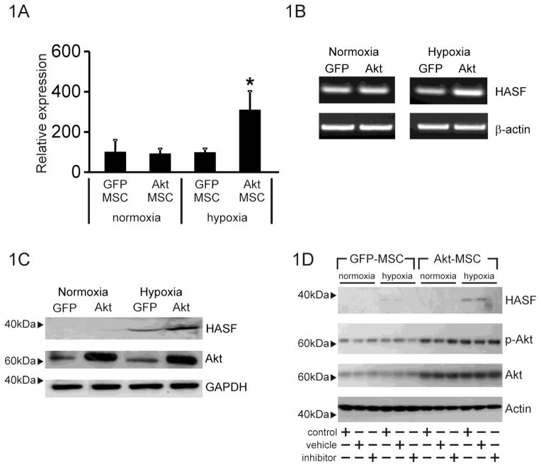
Figure 1. HASF is secreted by MSCs.
(A) Affymetrix microarray expression data of HASF in Akt-MSCs and control GFP-MSCs under normoxic or hypoxic conditions for 6hr. * P<0.001. Values are mean ± SE (n=3). (B) RT-PCR validation of mouse HASF expression in Akt-MSCs (Akt) and control GFP-MSCs (GFP) under normoxic or hypoxic conditions for 6hr. Beta-actin was used as internal control. (n=3). (C) Immunoblot analysis for HASF expression in conditioned media from MSCs using an anti-HASF polyclonal antibody. GFP: control GFP-MSCs, Akt: Akt-MSCs. Akt and GAPDH levels in the cell lysates served as controls. (D) Immunoblot analysis of conditioned media for HASF expression from MSCs cultured under normoxic or hypoxic conditions in the presence of the secretion inhibitor brefeldin-A (1μg/ml). Cell extracts (7.5μg) were probed with phospho-Akt, Akt and actin. Phospho-Akt and Akt to show that brefeldin-A had no effect on Akt activity or Akt expression, actin served as a loading control (n=3).

