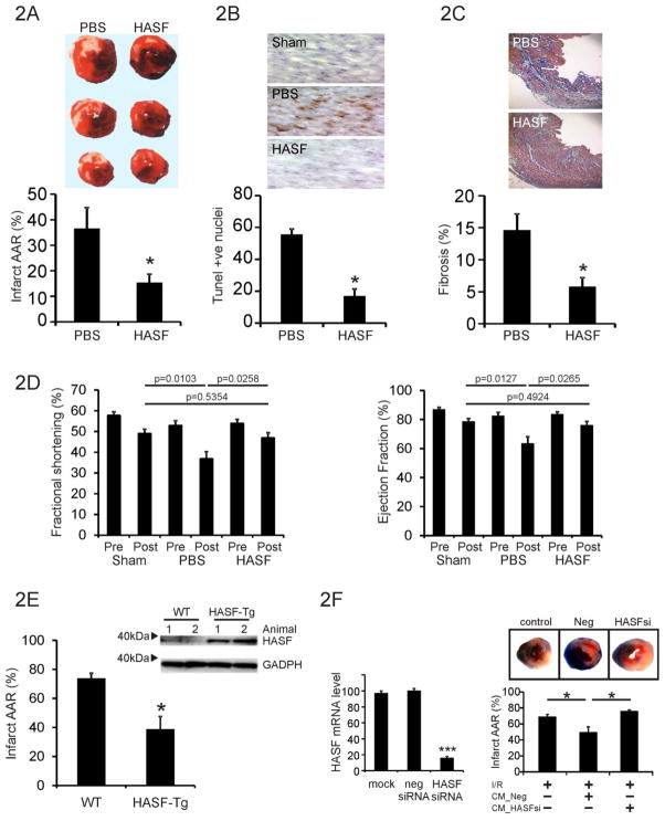
Figure 2. HASF protects the heart against myocardial injury.
(A) Quantification of infarct size (% of Infract area/AAR) in the rat hearts undergone I/R and randomly divided into PBS control and HASF injected groups. Data are presented as means ± SE of replicates, n= 10. Representative photos from each group are also shown. * P<0.001. (B) TUNEL staining in control or HASF injected animals from parallel experiments as above. The means ± SE of replicate heart samples per group are presented, n= 10, * P<0.001. Sections were counterstained with hematoxylin. Representative photos from each group are also shown. (C) Analysis of fibrosis 4 weeks after the initial ischemia/reperfusion injury. Percentage (%) of fibrosis was calculated as collagen positive area/total area. Data represent mean ± SE, n = 6–8 mice per group, * P<0.001. Representative photos from each group are also shown. (D) Echocardiograph data of rats following HASF injection, fractional shortening and ejection fraction are shown. P-values as indicated. Sham n=8, PBS n=9, HASF n=10. (E) Immunoblot assay of left ventricle protein lysates in Tg and WT mice. Antibodies against HASF were used. GAPDH served as loading control. Hearts from wild type (WT) and alpha-MHC driven human HASF transgenic mice (α-MHC HASF-Tg) were analyzed by TTC staining after Ischemia/reperfusion injury. AAR was equal on all groups tested (Supplementary Figure 2B). *, P < 0.05, Data are presented as means ± SE of replicates, n= 4–5. (F) Left panel: Akt-MSCs were transfected with lipid reagent alone (mock), negative control siRNA (neg siRNA), and HASF siRNA (HASF siRNA). qPCR was used to determine HASF RNA expression. Conditioned media was collected from cells transfected with either negative control or HASF siRNA. Right panel: TTC staining showing damage by Ischemia/Reperfusion (I/R) in mice hearts treated with conditioned media from Akt-MSCs. (% Infract area/Area at risk (AAR). CM_Neg = conditioned media from MSCs treated with scrambled negative control siRNA; CM_HASF-si = conditioned media from MSCs treated with SiRNA targeting HASF; AAR was equal on all groups tested (Supplementary Figure 2D).* P < 0.05, Data are presented as means ± SE of replicates, n= 10.

