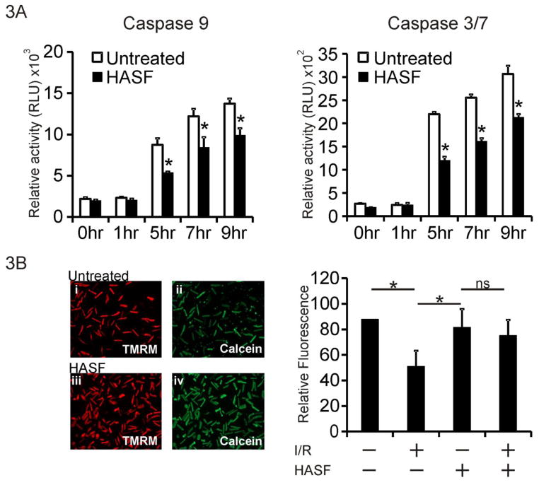
Figure 3. HASF protects cardiomyocytes against cell death in vitro.
(A) Caspase elisa assay in adult rat cardiomyocytes treated with 10 nM HASF, or PBS control for 30 minutes, and then challenged with 100 μM of H2O2 for various time lengths. Data are presented as means ± SE of replicates, * P<0.05, n=4–6. (B) Left panel: Representative images of cardiomyocytes subjected to 3hr hypoxia, 60 minutes reoxygenation (I/R) showing cells pretreated with HASF and control PBS treated cells. Calcein (Green); Mitochondrial dye TMRM (red). Increased Green fluorescence acts a surrogate marker for increased inhibition of mPTP channel opening. Right panel: Quantitative fluorescence analysis of images shown in left. Inhibition of mPTP channel opening was defined as the percentage of Calcein intensity (green fluorescence) normalized to viable cell number. Data are presented as means ± SE of values estimated from 8 images per condition.

