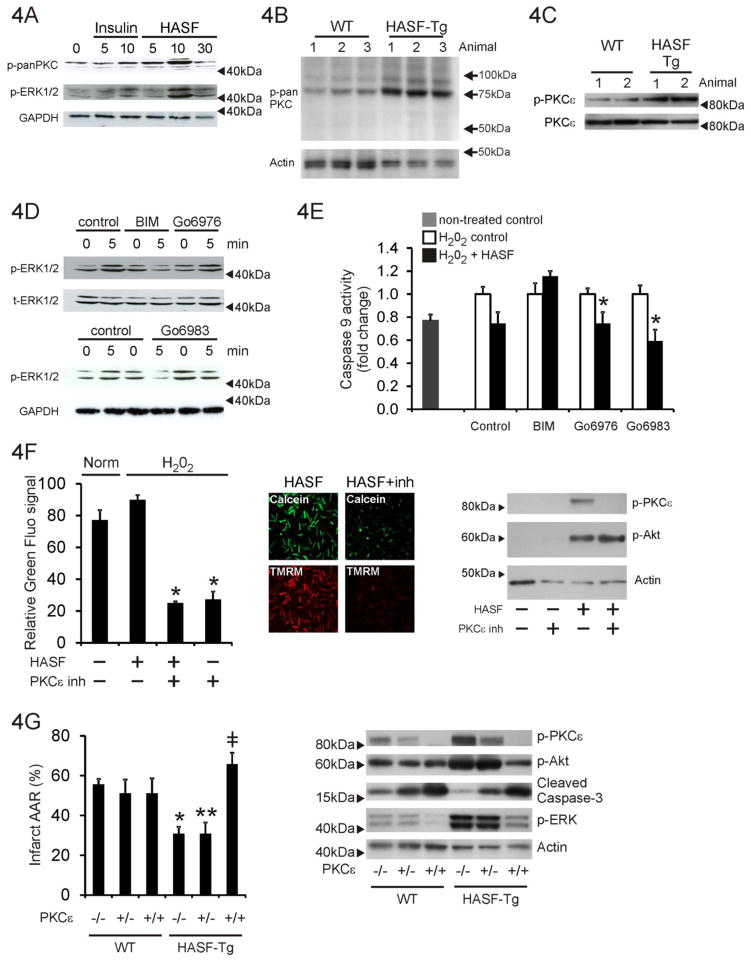
Figure 4. The cytoprotective effects of HASF are mediated by PKCε.
(A) Immunoblot analysis for PKC and ERK protein phosphorylation levels in serum-restricted adult rat cardiomyocytes stimulated with HASF recombinant protein for various times. Insulin was used as a positive control. min: minutes of treatment (B) Immunoblot analysis of left ventricle protein lysates in wild-type and HASF-Tg mice for pan-phospho-PKC (PDK1 phosphorylation motif). Actin was used as a loading control (C) Immunoblot assay of left ventricle protein lysates in Tg and WT mice. Antibodies against PKCε phosphorylation (S729) and total PKCε were used. GAPDH was used as a loading control. (D) Adult rat cardiomyocytes were pre-treated with 3 μmol/L PKC inhibitors BIM, Gö6976, or Gö6983 for 30 minutes, stimulated with 100nmol/L HASF recombinant protein for 5–10 minutes and harvested. Total cell lysates were used to evaluate the levels of ERK1/2 phosphorylation by immunoblot blot analysis. GAPDH was used as a loading control. (E) Serum-restricted adult rat cardiomyocytes were treated with 3 μmol/L PKC inhibitors or vehicle DMSO for 30 minutes, followed by HASF recombinant protein or PBS control treatment for 30 minutes and treatment with 200μmol/L H2O2 for 3 h. Apoptosis was determined by detection of caspase-9 activity using a luminescent assay. All samples were measured in triplicates and the data were normalized to the corresponding non HASF- control cells. *P < 0.05 vs. corresponding PBS treated control cells. Note that PKCε is the only isoform differentially affected by BIM (see also Supplementary Table 3). (F) Quantitative image analysis of mPTP channel opening during hypoxia/reoxygenation as described above in the presence of a peptide PKCε inhibitor. Fluorescence levels are shown in the graph. Data are shown as means ± SE of values estimated from 8 images per condition. * P<0.01. Representative images are shown on the left. Protein extracts (20μg) were immunoblotted for phospho-PKCε, phospho-Akt and actin. (G) HASF mediated cardioprotection is abrogated in mice that lack PKCε. HASF Tg mice were crossed with PKCε knock-out mice. Groups of mice from all the resulting genotypes were then subjected to ischemia/reperfusion injury and TTC staining as described before. The calculations of % of Infract/AAR are presented for each group. AAR was equal on all groups tested (Supplementary Figure 4B). Only statistical significant effects are highlighted. * P < 0.05, HASF Tg vs. WT; ** P <=0.05 HASF Tg: PKCε+/− vs. PKCε+/− control; #, P < 0.01 HASF Tg: PKCε−/− vs. HASF Tg: PKCε+/− or vs. HASF Tg: PKCε+/+. Data are presented as means ± SE of replicates, n= 4–5. Protein extracts (20μg) were immunoblotted for phospho-PKCε, phospho-Akt, cleaved caspase-3, phospho-ERK and actin.

