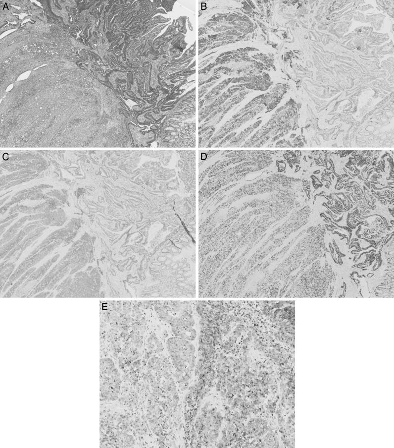
Fig. 3.
(A) The tumor was composed of high-grade large-cell endocrine cell carcinoma with moderately differentiated tubular adenocarcinoma. Colon: HE (×40). (B) Chromogranin A (+). Colon: chromogranin A (×40). (C) Synaptophysin (+). Colon: synaptophysin (×40). (D) The Ki-67 labeling index was high in both lesion (80%). Colon: Ki-67 (×40). (E) The tumor in the liver was also composed of high-grade large-cell endocrine cell carcinoma with moderately differentiated tubular adenocarcinoma. Liver: chromogranin A (×200).

