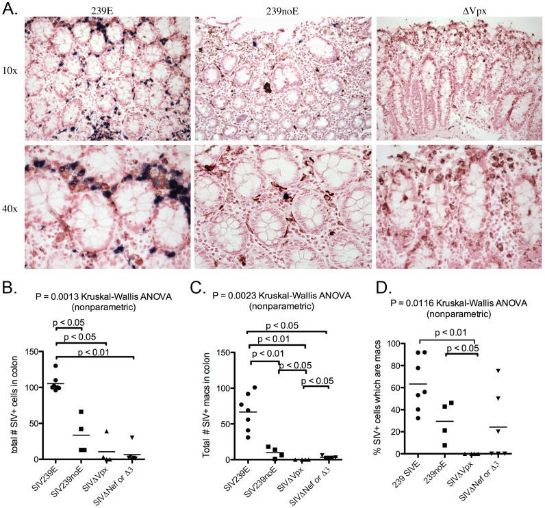
Figure 5. Double-label immunohistochemistry and in situ hybridization of colon.
(A). Representative images of SIV ISH (blue) with double-label immunohistochemistry for macrophage marker Ham56 (DAB, brown) in rhesus macaques in groups SIV239E (left), SIV239noE (middle), and SIVΔvpx (right). SIV+ Ham56+ macrophages were frequent in SIV239E monkeys (left), but absent in the SIVΔvpx group (right). (B). SIVΔvpx-infected monkeys had significantly less virus in the colon (mean 10.5 SIV+ cells) compared to monkeys infected with SIV239 (SIV239E mean 105.3 SIV+ cells, SIV239noE mean 33.5 SIV+ cells), but no difference with animals in the SIVΔnef/SIVΔ3 group. (C). SIVΔvpx monkeys (mean 0 cells) had significantly fewer SIV+ Ham56+ macrophages compared to SIV239E (mean 66.7 cells), SIV239noE (mean 9.8 cells), and SIVΔnef/SIVΔ3 groups (mean 3.3 cells) groups. (D). There was a significantly lower percentage of SIV+ cells that were macrophages in SIVΔvpx-infected rhesus (mean 0%) compared to both the SIV239E (mean 63.3%) and SIV239noE (mean 22.4%) groups and a trend in the SIVΔnef/SIVΔ3 group (mean 25.6%).

