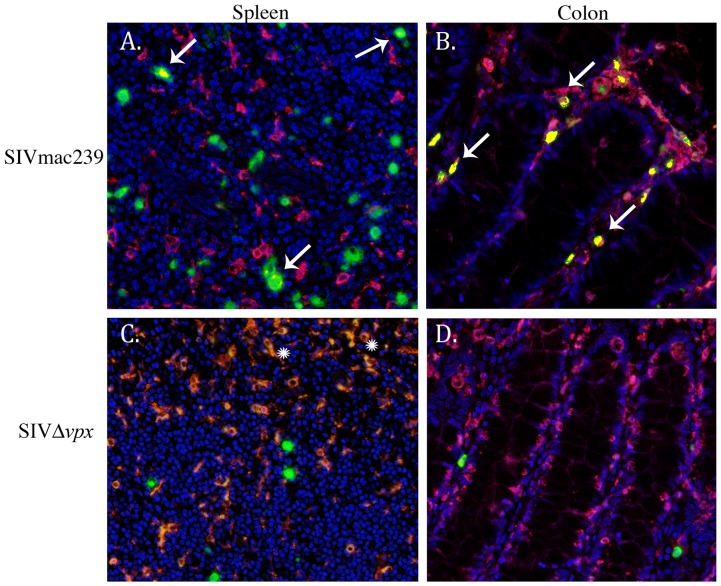
Figure 6. Spectral imaging and colocalization of double-label immunohistochemistry slides of spleen and colon.
Immunofluorescence with SIV nucleic acid (green), Ham56+ macrophages (red), and coexpression (yellow, arrows) demonstrate that infected macrophages are readily evident in SIVmac239-infected monkeys (A, B, arrows), particularly within the colon (B), but are absent in spleen and colon from SIVΔvpx-infected monkeys (C, D). Snowflake symbols in C denote autofluorescence in red pulp macrophages. Original magnification 40× (A–D).

