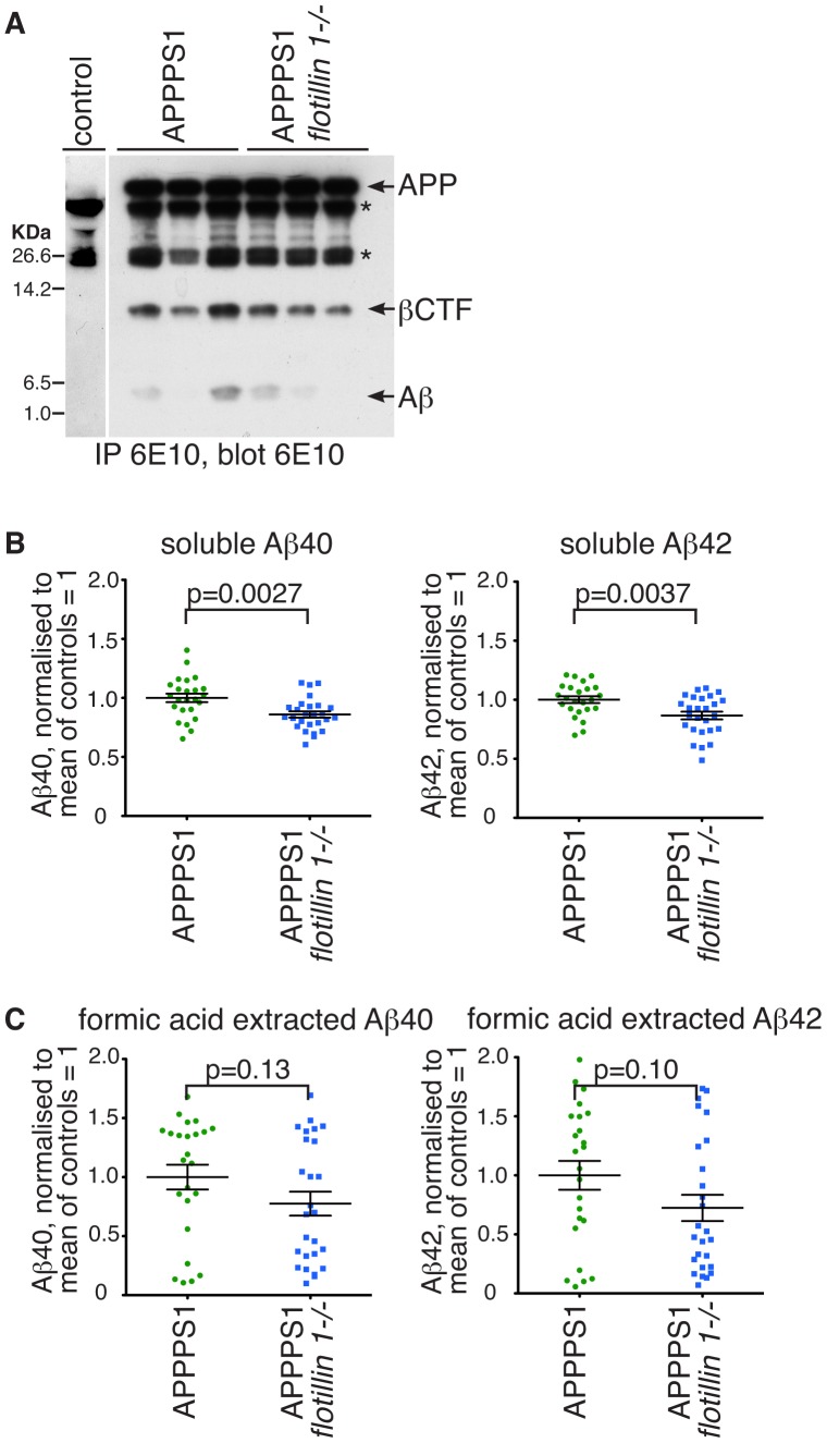
Figure 3. Deletion of flotillin 1 reduces the accumulation of both soluble Aβ, and Aβ in formic-acid extractable plaques, in brains of APPPS1 mice.
A. APP from RIPA buffer solubilised lysates of mouse brain was immunoprecipitated with the monoclonal antibody 6E10, and the precipitates analysed by Western blotting with the same antibody. The bands corresponding to full length APP (APP), β C-terminal fragment of APP (βCTF), and Aβ are indicated. Bands with an * are present in mice not expressing human APP, and are most likely antibody heavy and light chains. 3 mice of each genotype were analysed. Approximate positions of protein molecular weight markers are indicated. B. Brain tissues from 12 week old APPPS1 or APPPS1, flotillin 1-/- mice were harvested and Aβ levels were measured quantitatively using ELISA. Soluble Aβ40 and Aβ42 were present in the supernatant after tissue homogenisation and centrifugation at 20,000 rcf. Each data point represents assay from the brain of one mouse. Bars are SEM. C. Brain tissues were harvested as in B above, but Aβ40 and Aβ42 were extracted with 70% formic acid from the pellet, after tissue homogenisation and centrifugation, and the levels assayed using ELISA. Each data point represents assay from the brain of one mouse. P values were calculated using Student's t-test. Bars are SEM.

