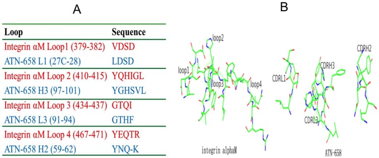
Figure 5. Sequence and 3-dimensional structural similarity between αM binding loops and ATN-658 CDR loops.

(A) Sequence alignment of integrin αM loops to ATN-658 CDR loops. (B) Similar spatial arrangement between integrin αM loops and the ATN-658 CDR loops. Structure of αM was a homology model built from the known integrin structures (http://prosite.expasy.org/cgi-bin/pdb/get-pdb.pl?1a8x).
