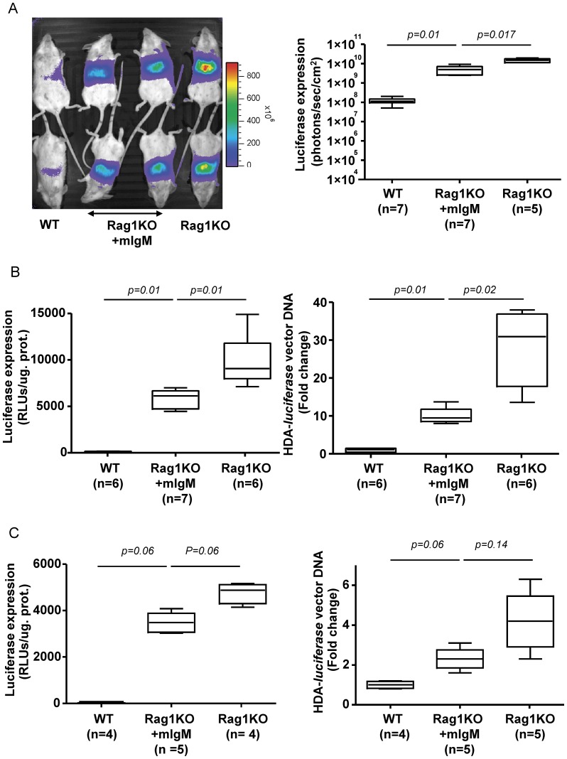
Figure 5. Monoclonal IgM of a specificity unrelated to adenovirus reduces liver gene transfer by HDA vectors.
A) Representative photographs of light emission from the liver region of mice intravenously injected with 5×10¹⁰ vp/mouse 48 h prior to bioluminescence (left panel). When indicated, Rag1KO were pre-injected twice with 130 µg of monoclonal IgM of irrelevant specificity. The dot graph on the right shows quantitative data from the same experiment. Experiments B and C were performed seven days following gene transfer with progressively reduced doses of HDA-luciferase (3.6×10¹⁰ vp/mouse and 2.2×10⁹ vp/mouse, respectively). Gene transfer was assessed by measuring luciferase activity ex-vivo in homogenates from liver samples (left panel). The relative liver content of vector DNA was measured by quantitative PCR (right panel). WT, Wild-type; Rag1KO, BALB/C Rag1−/− (C.129S7(B6)-Rag1tm1Mom/J; pIgM, polyclonal immunoglobulin M; mIgM, monoclonal immunoglobulin M, mIgG, monoclonal immunoglobulin G.; HDA-luciferase, helper-dependent adenoviral vectors encoding luciferase; RLU, Relative Light Unit. Experiments were performed twice.

