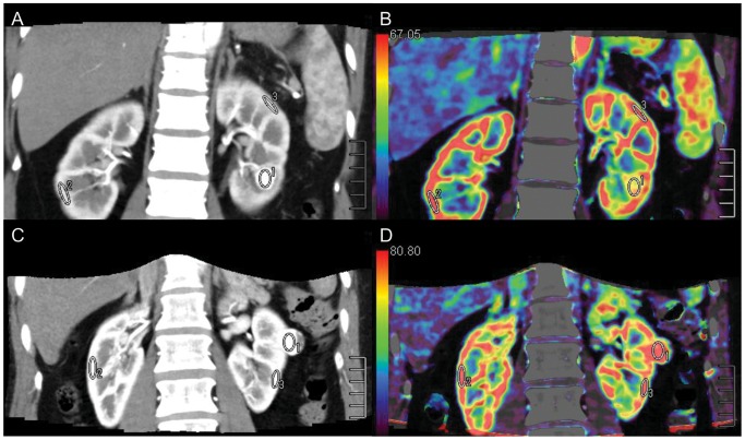
Figure 2. ROIs (ROI 1, ROI 2, and ROI 3) were placed in the tumor and in healthy ipsi- and contralateral renal cortex.
ROI 1 was the ROI of tumor tissue. AML with minimal fat (A) (mean CT value = 255.4 HU) with a sufficient blood supply had an abnormal enhancement pattern that was similar to clear cell RCC (C) (mean CT value = 251.4 HU), while Equiv BV was much lower in AML (B) (mean Equiv BV = 57.7) than in clear cell RCC (D) (mean Equiv BV = 85.2).

