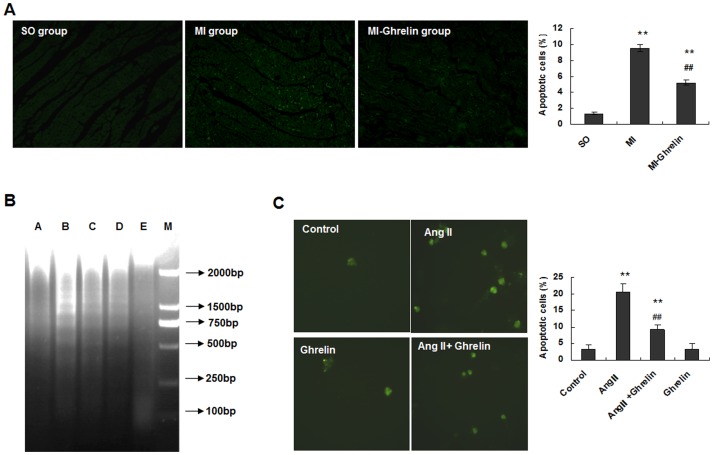
Figure 3. Ghrelin inhibited cardiomyocyte apoptosis both in vivo and in vitro.
(A) TUNEL analysis was performed after the end of the ghrelin treatment. The TUNEL-positive cells (apoptotic cells) are indicated by arrows. The data are presented as the means ± SD. **P<0.01 vs. SO group, ##P<0.01 vs. MI group. (B) Ang II-induced DNA fragmentation in cardiomyocytes with or without ghrelin. Cultured cardiomyocytes from neonatal rats were stimulated with or without Ang II and ghrelin for 24 hours. The cardiomyocyte lysate was first incubated with RNase and then with proteinase K. By this method, only fragmented DNA was extracted. The DNA was separated by electrophoresis on a 1.5% agarose gel and stained with ethidium bromide. Lane A, vehicle-treated cardiomyocytes; lane B, cardiomyocytes incubated with 0.1 µmol/L Ang II; lane C, cardiomyocytes incubated with 1 µmol/L Ang II; lane D, cardiomyocytes incubated with 0.1 µmol/L ghrelin and 0.1 µmol/L Ang II; lane E, cardiomyocytes incubated with 0.1 µmol/L ghrelin. (C) Apoptosis of cardiomyocytes treated with Ang II and ghrelin. Cardiomyocytes were incubated in culture medium (control), 0.1 µmol/L Ang II, 0.1 µmol/L ghrelin, or 0.1 µmol/L ghrelin + 0.1 µmol/L Ang II for 24 hours. Magnification: ×200. The graph presents the percentage of TUNEL-positive cells determined from 100 cells in 3 independent experiments. The data are presented as the means ± SD. **P<0.01 vs. the control group; ##P<0.01 vs. the Ang II group.

