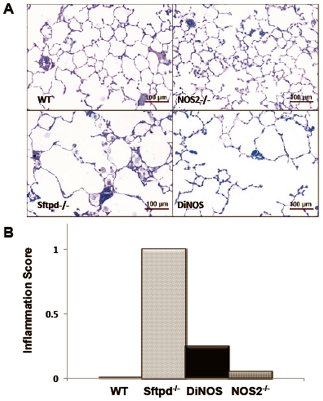
Figure 2. Lung histology reveals parenchymal inflammation in Sftpd−/− mice and reduced enlargement of distal airspaces in DiNOS.

(A) Representative micrographs of lung sections from WT (upper left), NOS2−/− (upper right), Sftpd−/− (lower left) and DiNOS (lower right) mice (toluidine blue staining). Normal lung architecture can be seen in WT and NOS2−/− mice. In Sftpd−/− the distal airspaces are enlarged and filled with foamy appearing alveolar macrophages. Enlargement of distal airspaces appears to be less pronounced in DiNOS compared to Sftpd−/− mice. (B) Histological scoring of lung inflammation. Median inflammation scores were determined by blinded evaluation of stained sections from each genotype group as described in Online Supplement Methods. Data shown are Median, n = 7 animals per genotype.
