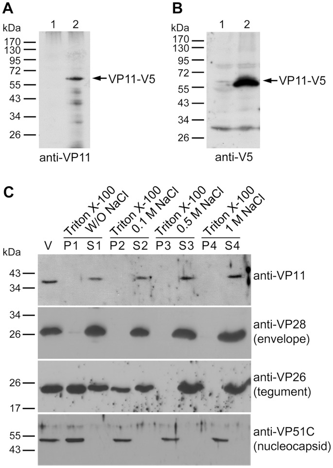
Figure 2. Determination of VP11’s location in the WSSV virion.
Western blot analysis of the full-length recombinant WSSV VP11 (VP11-V5) expressed in Sf9 cells using either (A) anti-VP11 antibody or (B) anti-V5 antibody. Lane 1: cell lysate of pDHsp/V5-His transfected Sf9 cells. Lane 2: cell lysate of pDHsp/VP11-V5-His transfected Sf9 cells. (C) Intact WSSV virions were subjected to detergent and NaCl treatment as indicated. After fractionation, the pellet (P) and supernatant (S) fractions were separated on SDS-PAGE and detected by Western blotting to produce profiles that are characteristic of envelope, tegument and nucleocapsid proteins (6). Three representative WSSV structural proteins are shown for comparison. Lane V is the untreated purified virus.

