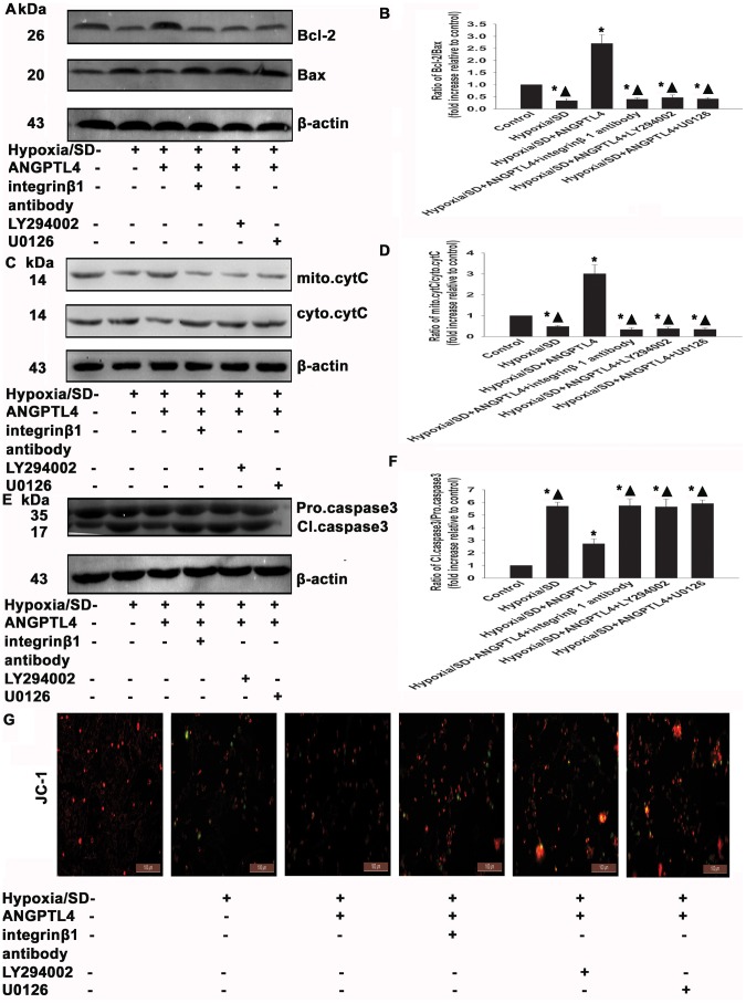
Figure 10. ANGPTL4 exerted anti-apoptotic effects via inhibition of mitochondrial dysfunction.
MSCs were treated with hypoxia/SD for 24 hr. In parallel experiments, cells were pretreated with either integrin β1 antibody (20 µM) or LY294002 (25 µM) or U0126 (20 µM) for 90 min before exposure to hypoxia/SD. When present, ANGPTL4 (100 ng/mL) was added in the presence of each drug for 1 hr prior to exposure to hypoxia/SD. All drugs were maintained in the incubation medium throughout the hypoxia/SD treatment period. Bcl-2/Bax ratio (A and B), cytochrome C (C and D), and caspase-3 (E and F) were detected by Western blotting. (Each example shown is representative of three experiments). Fold changes were compared to control. (G) MMP with JC-1 staining. (Each column represents the mean ± SD of three independent experiments; *P<0.05 vs. control; ▴P<0.05 vs. hypoxia/SD+ANGPTL4 −100 ng/mL).

