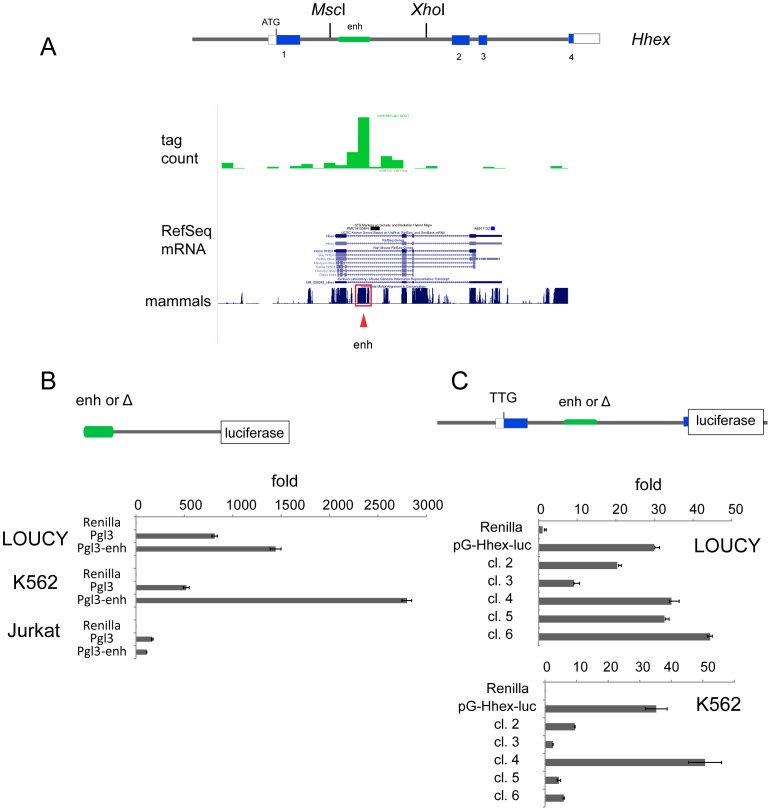
Figure 5. T-ALL expression of HHEX requires its promoter and enhancer.
(A) Schematic shows the structure of the mouse Hhex gene with 4 exons (coding sequences shown in blue). We analyzed ChIP-seq data from anti-Ldb1 that showed occupancy within intron 1 (green bar graph). The RefSeq mRNAs for Hhex are shown below the sequencing tag counts. The bar graph under this shows the conservation across multiple mammalian species. The red box denotes an enhancer that was previously functionally characterized as specifying blood-specific expression of Hhex. (B) We cloned the enhancer shown in panel A 5′ to the SV40 promoter and transfected it into LOUCY, K562, and Jurkat cell lines along with pCMV-Renilla luciferase vector. Luciferase values were normalized to Renilla to correct for transfection efficiency and expressed as fold over Renilla alone. (C) We constructed a luciferase expression vector with 1 kb of the Hhex promoter, exon 1, intron 1, and replaced exon 2 with the luciferase cDNA. We transfected this reporter (pG-Hhex-luc) into K562 and LOUCY cells. Clones 1–6 have 50–100 bp deletions 5′ to 3′ within the enhancer region. Luciferase expression was normalized to Renilla for transfection efficiency and expressed as fold above that seen in Renilla alone.

