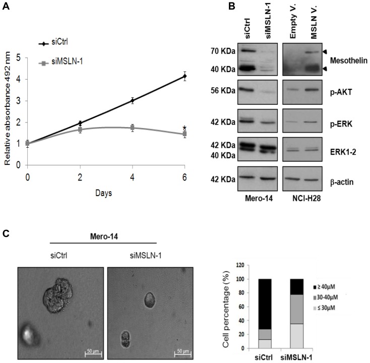
Figure 2. Role of MSLN in cellular growth.
A. SRB proliferation assay in Mero-14 cells treated with 40 nM of the siCtrl or siMSLN-1 (*P = <10−4). Error bars represent SEM of three independent experiments, each performed in quadruplicate. B. Western blotting analysis of MSLN, p-AKT, p-ERK, and ERK1-2, on Mero-14 cells treated with siCtrl, or siMSLN-1, and on NCI-H28 cells transfected with an empty vector (pcDNA3.1) or a plasmid overexpressing MSLN (pcDNA3.1-MSLN). β-actin was used as reference. The protein levels were confirmed with three independent experiments. C . The picture represents the phase contrast microscopy of Mero-14 cells cultured in 3D Matrigel-overlay chambers after silencing of MSLN (siMSLN-1 at 40 nM). Magnification 10X. Two different experiments were performed, each in triplicate. The percentages of Mero-14 cells classified according to the dimensions of the spheres following treatments with siCtrl or siMSLN-1 in 3D Matrigel-overlay chambers were also reported. Legend to figure 2: Gray line = cells treated with siMSLN-1; Dark line = cells treated with siCtrl.

