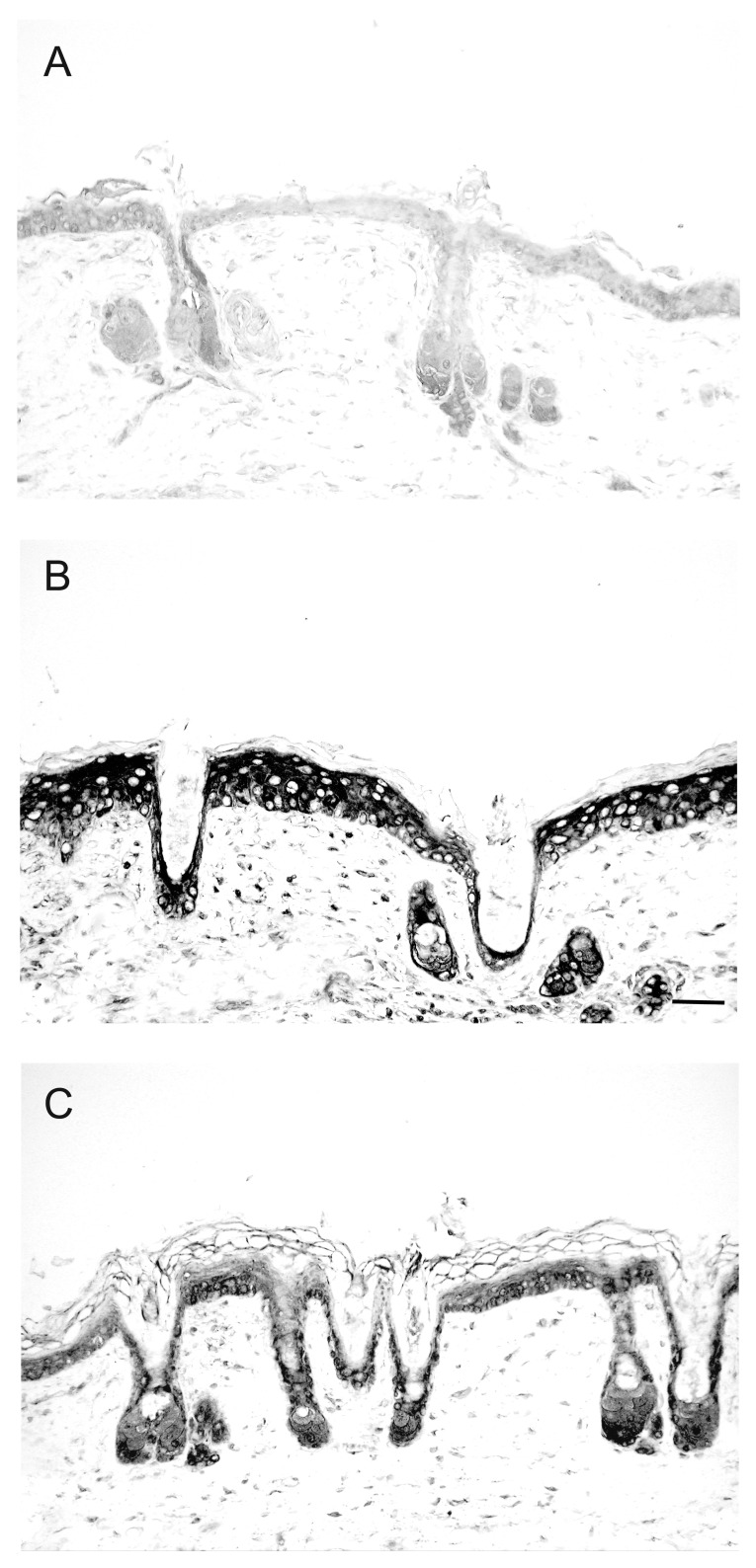
Figure 3. Reduction of UV-induced IL-6 expression by topical application of 1,25(OH)2D3 in mouse skin. Immunohistochemical detection of IL-6 in Skh:hr1 hairless mice skin was with a monoclonal antibody to IL-6 and a biotinylated secondary rabbit anti-goat IgG. Figures are representative dorsal skin sections (A) non-irradiated skin or (B) after solar simulated radiation followed by 48-h treatment with vehicle or (C) after solar simulated radiation followed by 48-h treatment with 1,25D (22.8 pmol/cm2). Reprinted from The Journal of Steroid Biochemistry and Molecular Biology. R.S. Mason, V.B. Sequeira, K.M. Dixon, C. Gordon-Thomson, K. Pobre, A. Dilley, M.T. Mizwicki, A.W. Norman, D. Feldman, G.M. Halliday, V.E. Reeve (2010).

An official website of the United States government
Here's how you know
Official websites use .gov
A
.gov website belongs to an official
government organization in the United States.
Secure .gov websites use HTTPS
A lock (
) or https:// means you've safely
connected to the .gov website. Share sensitive
information only on official, secure websites.
