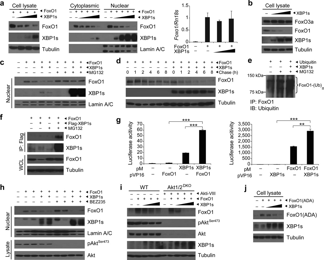
Figure 1.
XBP1s binds to FoxO1 and promotes its degradation. (a) XBP1s and FoxO1 protein levels in MEFs expressing FoxO1 and increasing levels of XBP1s. Foxo1 mRNA levels were analyzed by qPCR. (b) Endogenous FoxO3a and FoxO1 protein levels in MEFs expressing increasing levels of XBP1s. (c) Nuclear FoxO1 and XBP1s protein levels in MEFs treated with DMSO or MG132. (d) Pulse-chase analysis of FoxO1 stability in MEFs overexpressing XBP1s. (e) Ubiquitinylated FoxO1 levels in MEFs expressing ubiquitin and XBP1s after DMSO or MG132 treatment. (f) Immunoblotting of FoxO1 and XBP1s in XBP1s–immunoprecipitates from MG132-treated MEFs, expressing FoxO1 and XBP1s. Total FoxO1 protein level were determined in whole cell lysates (WCL). (g) Mammalian two-hybrid assay with pM-XBP1s and pVP16-FoxO1 or pM-FoxO1 and pVP16-XBP1s. (h) Nuclear FoxO1 and XBP1s protein levels in MEFs treated with 1 µM BEZ235 for 4 h. Phospho-AktSer473 and total Akt levels were determined in cell lysates. (i) FoxO1, pAktSer473, total Akt and XBP1s levels in WT and Akt1/2 double knockout (Akt1/2-DKO) MEFs treated with DMSO or 20 µM Akt inhibitor for 30 min. (j) Phosphorylation resistant mutant FoxO1(ADA) protein levels in MEFs expressing FoxO1(ADA) and increasing levels of XBP1s. Each experiment was independently reproduced three times. Error bars are ± s.e.m.; P values were determined by Student’s t-test (** P < 0.01, *** P < 0.001).

