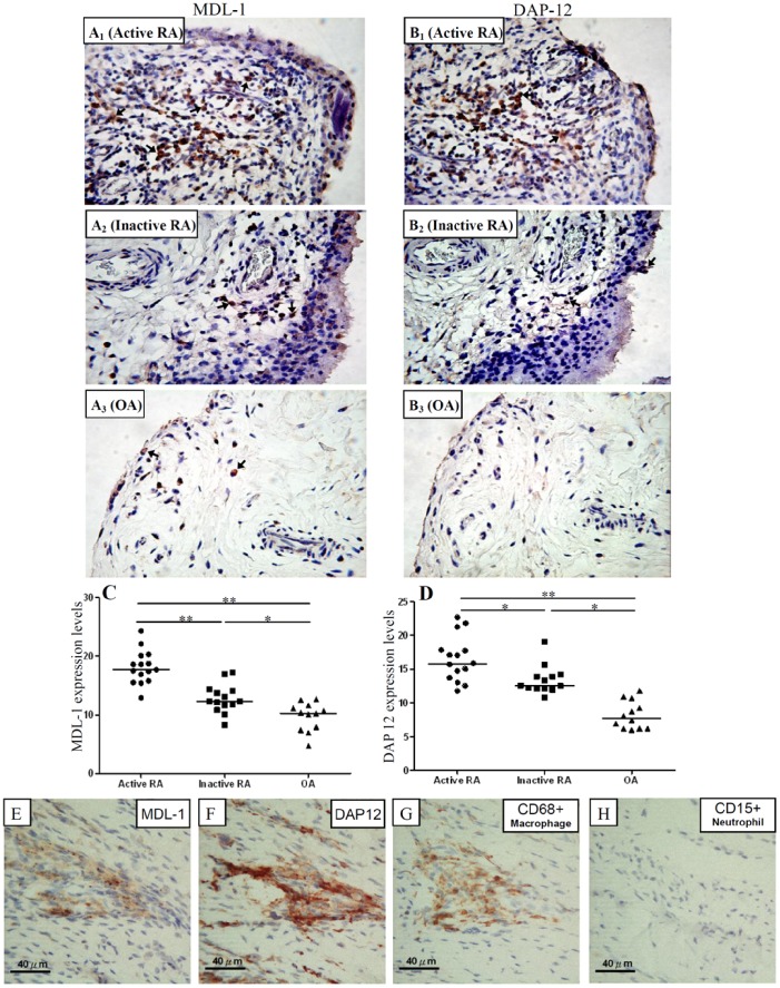
Figure 3. Representative examples of immunostaining with MDL-1 and DAP12 (arrows, original magnification x 200) obtained from synovial membranes of one active RA patient (A1 and B1; respectively), one inactive RA patient (A2 and B2; respectively), one OA patient (A3 and B3; respectively).
Comparisons of expression levels of MDL-1 and DAP12 on synovial membranes (C and D; respectively) are shown for 15 active RA patients, 14 inactive RA patients, and 12 OA patients. The horizontal line indicates median value for each group. *p<0.005, **p<0.001, as determined by Mann-Whitney U test. We also demonstrated that strongly positive staining of MDL-1 and DAP12 were expressed on CD68+ macrophages, not on CD15+ neutrophils in serial sections of synovial biopsy specimens from one RA patient (Figures 3E–3H).

