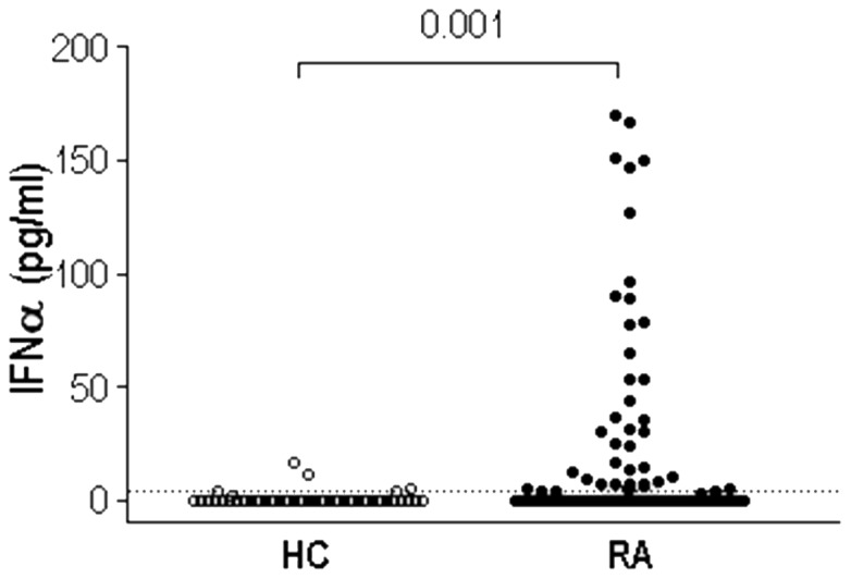
Figure 2. IFNα serum levels are increased in a subgroup of RA patients.

IFNα was quantified in 52 HC and 120 RA patients by CBA immunoassay. Dotted line represents HC 90th Percentile (4.092 pg/ml), used to classify RA patients in IFNlow (n = 80, 66%) or IFNhigh (n = 40, 33%). Differences were measured by Mann-Withney U-test.
