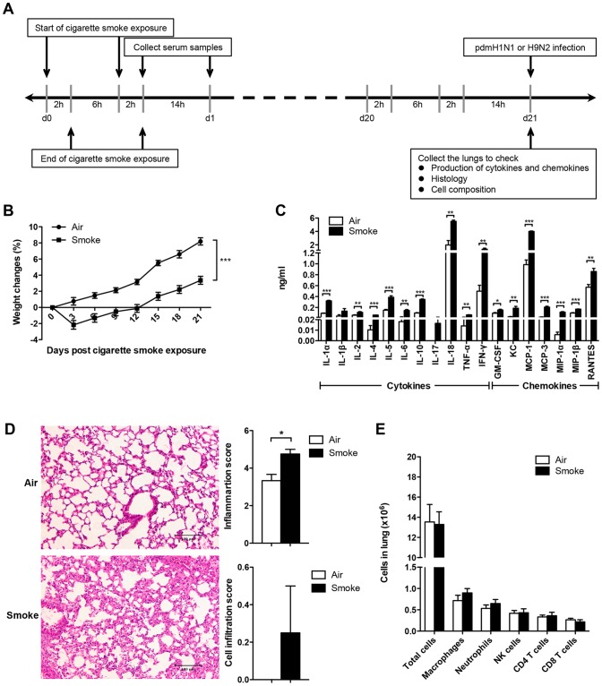
Figure 1. Cigarette smoke exposure alone up-regulated the inflammation of mice.
A) Study design for cigarette smoke exposure model. The mice were exposed to 4% (vol/vol, smoke/air) cigarette smoke for 2 hours per episode, 2 episodes per day for 21 days and then infected with pdmH1N1 or H9N2 virus. B) Body weight changes of the mice exposed to room air or cigarette smoke for 21 days. There were 50 mice per group. Data are representative of three independent experiments. C) Production of cytokines and chemokines in lung homogenates on day 21 of air or cigarette smoke exposed mice (fourteen hours after the last cigarette smoke exposure). Data are representative of three independent experiments. D) Histopathological analysis of pulmonary tissues collected on day 21 of air or cigarette smoke exposure (fourteen hours after the last cigarette smoke exposure). Results are representative pictures (200X) of hematoxylin and eosin stained pulmonary tissues. Scale bar: 100 µm. Inflammation score and cell infiltration score were evaluated by a board-certified pathologist. E) Absolute number of lung cells on day 21 of air or cigarette smoke exposure (fourteen hours after the last cigarette smoke exposure). Macrophages (CD11b+, F4/80+), neutrophils (CD11b+, Ly-6G+), NK cells (CD3−, NK1.1+), CD4+ T cells (CD3+, CD4+) and CD8+ T cells (CD3+, CD8a+). There were 4 mice per group (C, D, and E). Results represent mean ± SEM. *p<0.05; **p<0.01; ***p<0.001.

