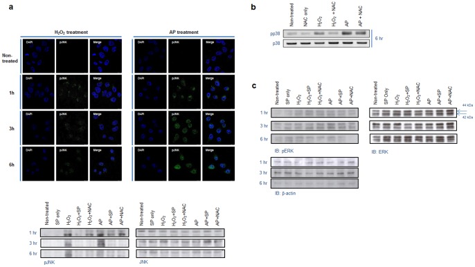
Figure 4. Air plasma-induced phosphorylation of JNK and p38.
(a) Phosphorylation of JNK following treatment with air plasma jet or H2O2 was analyzed via immunofluorescence staining and immunoblotting using anti-phospho-JNK antibody. The equivalent amount of total JNK proteins is shown as a quantitative loading control for JNK phosphorylation. (b) Phosphorylation of p38 induced by air plasma was visualized by immunoblotting using anti-phospho-p38 antibody. The comparable total p38 proteins are shown as a loading control for p38 phosphorylation. (c) Variation of phosphorylation status of ERK was not detected following treatment with air plasma or H2O2.

