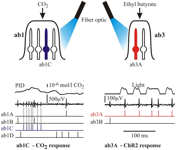
Figure 2. Three methods were used to stimulate basiconic sensilla.
Upper: CO2 responses were recorded from ab1C neurons using the apparatus of Fig. 1. Ethyl butyrate stimulation of ab3A neurons used the same apparatus with a filter paper cartridge delivering the odorant into the air/propylene stream. Optical stimulation of ab1C and ab3A neurons containing channelrhodoposin-2 was performed by a high intensity blue light emitting diode via a fiber optic. Lower: Multiple action potential recordings were separated by cluster analysis. Raw recordings from an ab1 sensillum to CO2 (left) and ab3 sensillum to light (right) are shown together with the inputs (PID and light traces), and the separated action potential times.

