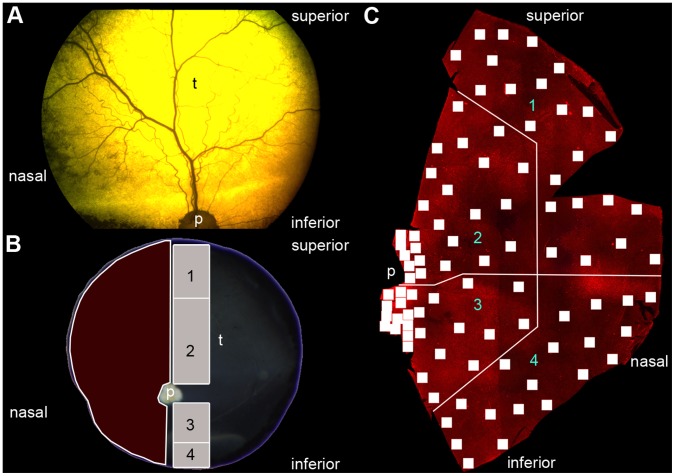
Figure 1. Orientation in the canine eye.
(A) Funduscopy of a RPE65−/− dog. (B) Extracted eye after euthanasia. The superior part of the eye determines the tapetum lucidum (t). The red marked area corresponds to the retinal flatmount used in the cone opsin expression analysis. The grey marked areas are used for cyrosections used in the bipolar cell sprouting analysis and the numbers correspond to regions shown in Figure 3 und 4. (C) Retinal flatmount for cone analysis. Every single quadrat stands for one microscope image (see material and methods). The retina was divided into 4 different regions. The numbers correspond to the regions shown in Figure 2. p = papilla, t = tapetum lucidum.

