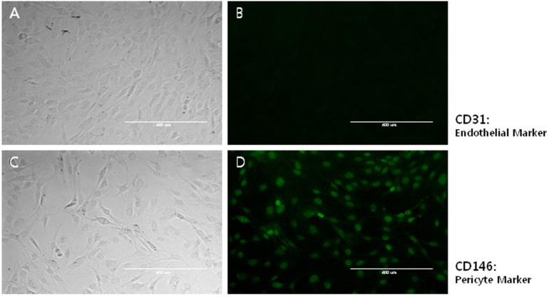
Fig. 1.
Characterization of primary mouse brain pericyte cultures. Primary mouse brain pericyte cultures were established as described in Materials and Methods. (A & B) Phase-contrast (A) or anti-CD31-labeled (endothelial marker protein) (B) images of the same pericyte cultures at passage 10. Scale bar=400 µm. Note that endothelial marker-positive cells were not detected. (C, D) Phase-contrast (C) or anti-CD146-labeled (pericyte marker protein) (D) pictures of the same pericyte cultures at passage 10. Scale bar=400 µm. At passage 10, all cells in the cultures were CD146-positive pericytes.

