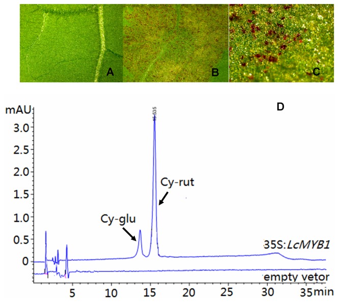
Figure 5. Color development in Nicotiana tabacum leaves following transient transformation.
Microscopic images showing anthocyanin accumulation in tobacco leaf infiltrated with: A) an empty vector (1× magnification) or B–C) 35S:LcMYB1 (8× magnification). D) Anthocyanin HPLC profiles of 35S:LcMYB1 extracts from tobacco leaf (top line) and empty vector (bottom line). Peaks identified at 520 nm: cy-glu, cyanidin-3-glucoside; cy-rut, cyanidin-3-rutinoside.

