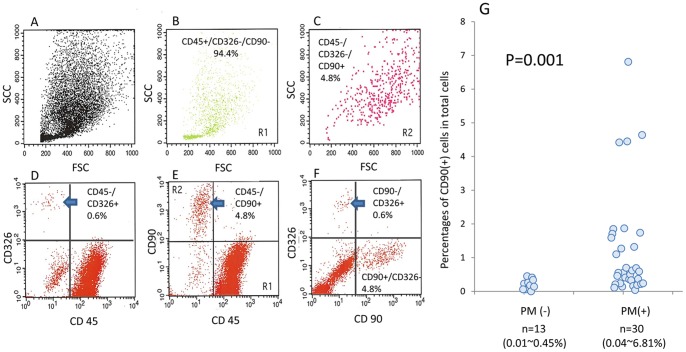
Figure 1. Intraperitoneal free cells were recovered from peritoneal lavages or ascites recovered at laparotomy from patients of gastrointestinal cancer and stained with FITC-conjugated anti-CD90, PE-conjugated anti-CD326 and PerCP-conjugated anti-CD45 and analyzed by FACS.
FACS profiles of a representative case were shown in A∼F. A: FSC/SCC, D: PerCP/PE, E: PerCP/FITC, F: FITC/PE. B and C show the FSC/SCC profiles of the cells located in Region 1 (CD45+, CD90−) and Region 2 (CD45−, CD90+), respectively. G: The cells were immunostained with FITC-conjugated anti-CD45 and PE-conjugated anti-CD90 mAbs and the ratio of CD45(−)CD90(+) cells were calculated in patients with or without peritoneal metastasis. The percentages of each cell population were calculated against total acquired cell counts of 104.

