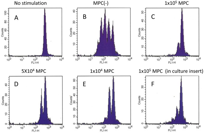
Figure 5. PBMC (1×10⁶) derived from healthy volunteers were stained with CFSE and cultured on plastic (A) or anti-CD3 coated (B∼F) plates in the presence or absence of the indicated number of MLCs for 4 days, and CFSE fluorescence intensities in CD3(+) T cells were analyzed with FACS.
In F, MLCs (1×10⁵) were added on culture inserts within the same well. Data shown is representative of results from 3 different experiments.

