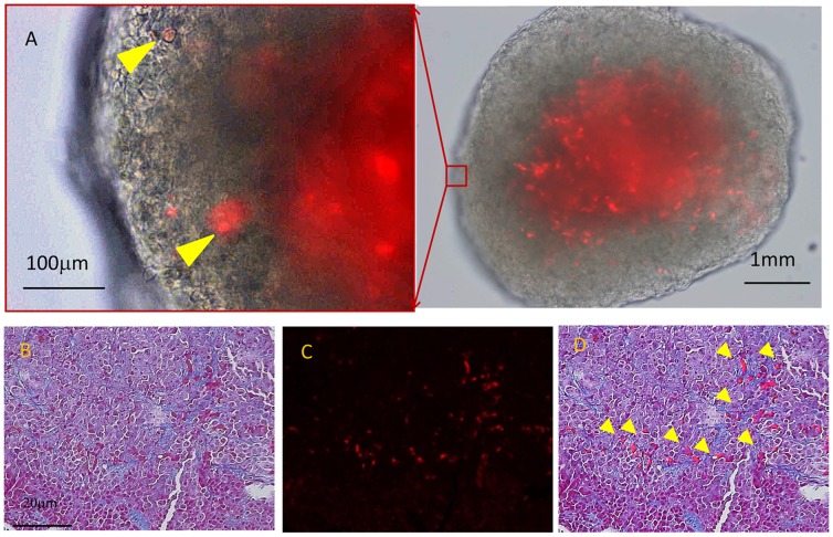
Figure 7. Peritoneal nodules developed in nude mouse after IP injection of MKN45 cells (1×10⁶) and PKH26-labelled MLCs (5×10⁵).
A: Nodules were excised and observed under fluorescence microscope. MLCs engrafted in metastatic nodules are highlight by arrow heads. Tissue sections of the peritoneal nodules were stained using the Masson-Trichrome method and observed under light (B) and fluorescence microscopy (C), and merged (D).

