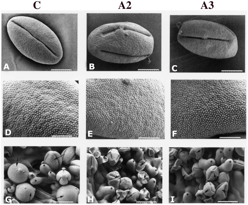
Figure 5. Ambient temperature SEM and low temperature SEM images of pollen in wildtype (panel C) and antisense (panels A2 & A3) plants.
A–C, ambient temperature low magnification of images of dehydrated pollen grains showing no significant difference in size or morphology (Scale bar = 9 µm). D–F, ambient temperature higher magnification images of the dehydrated pollen grains from all three lines showing no significant difference in the exine microstructure (Scale bar = 3 µm). G–I, low temperature SEM of selfed pollen, 2 hours after pollination. G, most wildtype pollen grains deposited on stigma are fully or partially hydrated; H and I, most pollen grains from antisense plants are not hydrated. G, arrow indicates pollen in hydrated condition or H and I, in non-hydrated condition. G, asterisk indicates stigmatic cell, and+indicates emerging pollen tube (Scale bar = 23 µm in G, and 30 µm in H and I).

