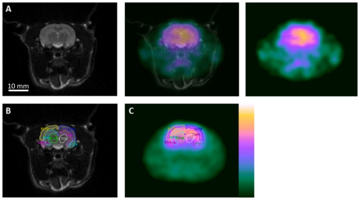
Figure 1. Coregistration and region of interest delineations for image analysis.
(A) Coregistration of [18F]-FMZ PET with T2 weighted MRI, (B) Delineation of regions of interest on MRI (RCTX right cortex, LCTX left cortex, RHPC right hippocampus, LHPC left hippocampus, RTHA right thalamus, LTHA left thalamus, RAmyg right amygdala, LAmyg left amygdala), (C) Application of regions of interest on to [18F]-FMZ PET image.

