
Fig. 2.
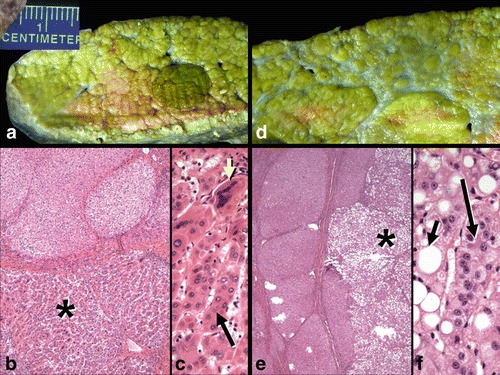
Explanted liver specimen from II.4 showing mixed micro-macronodular cirrhosis and several well-differentiated hepatocellular carcinomas. (a) A well-circumscribed carcinoma (dark green nodule) is present. (b) Microscopic section of the lesion seen in panel A shows an inactive cirrhosis at top and the hepatocellular carcinoma (*) below. (c) High magnification of the tumor in panel B shows partial microtrabecular pattern (long arrow) and many giant, multinucleated tumor cells (short yellow arrow). (d) Another nodule is grossly pale and is a steatotic hepatocellular carcinoma. (e) The underlying inactive cirrhosis (left) contrasts to the multilobulated fatty carcinoma at right (*). (f) The steatotic carcinoma shows many neoplastic hepatocytes with large fat vacuoles (short arrow) as well as focal microtrabecular growth (long arrow). (Hematoxylin and eosin stains; b: x 40; c: x 200; e: x 40; f: x 200)