Fig. 1.

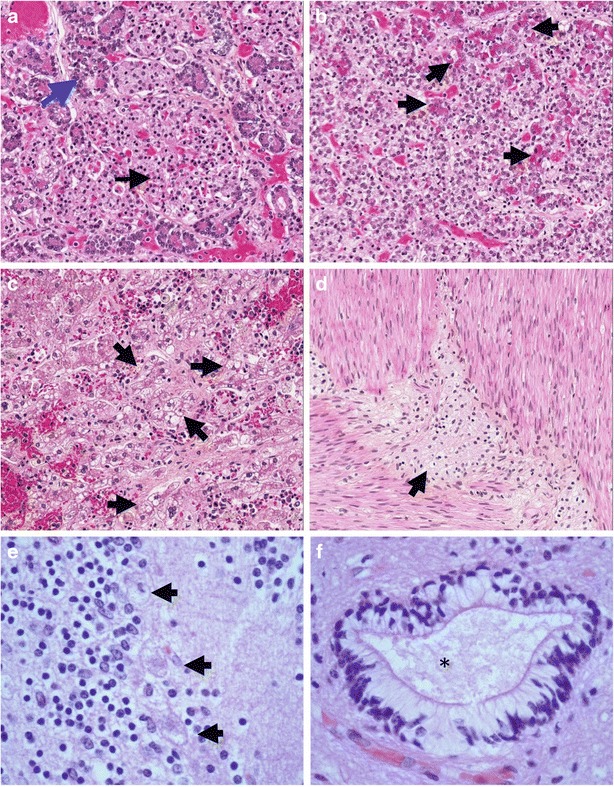
Pathologic findings in Patient 1. (a) Section through pancreas showing enlarged, vacuolated islet cells (black arrow), with relatively normal-appearing acinar cells (blue arrow) at periphery (b) Pituitary gland contains a majority of pale, vacuolated cells with a foamy appearance; some acidophilic cells with a superimposed foamy quality (arrows) are seen (c) Sections of liver show granular, swollen, pale, foamy cells throughout (some clusters indicated by arrows). The stored material does not stain by Oil Red-O or periodic acid-Schiff histochemical stains (data not shown) (d) Ganglion cells of the myenteric plexus (arrow) have enlarged, pale cytoplasm with a foamy quality (e) Cerebellar cortex: three Purkinje cells are swollen, with cytoplasmic accumulation of foamy storage material and loss of Nissl substance (arrows) (f) Ependymal canal, spinal cord: ependymal cells have a clear cytoplasm due to presence of storage material (asterisk indicates center of canal)
