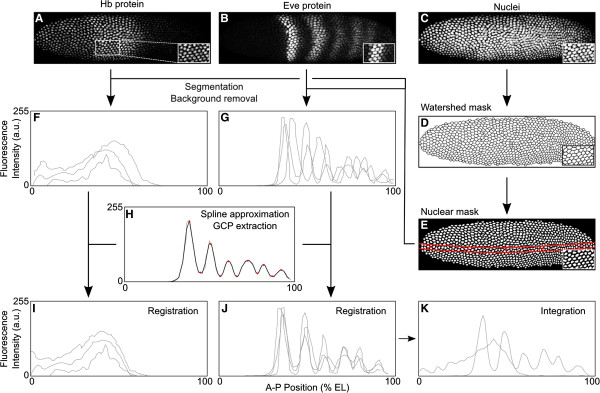
Figure 3.

Summary of image processing. Upper panels show scanning confocal microscopy images of an example embryo stained against Hb protein (A), against Eve protein (B), and with a nuclear counterstain (C). Insets show magnified details at the position indicated by a white square in (A). Nuclear counterstains (C) are used to generate binary watershed masks (D) and nuclear masks (E) for image segmentation. Individual expression profiles after image segmentation and background removal are shown for Hb (F) and Eve (G). These profiles are extracted from a 10% strip along the midline of the embryo, as shown by red lines in (E). Data registration is performed by approximating the Eve pattern using quadratic splines (H): Ground Control Points (GCPs, red dots in H) are extracted and used as the basis for an affine coordinate transformation which minimizes embryo-to-embryo variation in their position across the dataset (J). The same transformation is applied to the Hb profile (I). Finally, individual expression profiles belonging to the same time point are classified into 100 bins along the antero-posterior (A-P) axis, and data points within bin are averaged to yield an integrated dataset for both Hb and Eve (K). See Methods for details. a.u., arbitrary units; EL, egg length.
