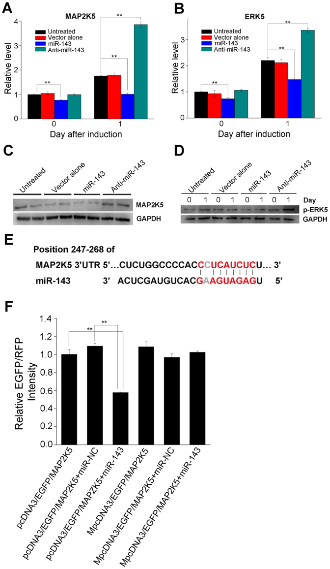
Figure 5. miR-143 regulates ADSC adipogenesis by inhibiting the MAP2K5–ERK5 signaling pathway.
(A) MAP2K5 mRNA levels were downregulated in cells infected with an miR-143-expressing lentiviral vector (miR-143 group) on days 0 and 1 following MDI induction. (B) As the direct downstream of MAP2K5, ERK5 mRNA levels were similarly downregulated in the miR-143 group. (C) MAP2K5 protein levels were evaluated by western blot analysis 1 day after incubation with MDI. (D) Phosphorylated ERK5 (p-ERK5) levels were determined by western blot analysis at the indicated time points after ADSCs were treated with MDI. (E) A sequence within the MAP2K5 mRNA that is complementary to miR-143 was identified using publicly available algorithms. (F) Cells were cotransfected with an EGFP reporter plasmid (pcDNA3–EGFP–MAP2K5) or the mutant vector (MpcDNA3–EGFP–MAP2K5) and the pDsRed–C1 plasmid, either alone or in combination with an miR-143 mimic. EGFP and RFP levels were measured 48 h after transduction with an F-4500 fluorescence spectrophotometer. The fluorescence value in the control group was set to 1. Histograms show the normalized mean ± SD fluorescence intensity of three independent experiments. All other data are shown as means ± SD. **P < 0.01 vs. untreated cells or cells transfected with the empty vector.

